---
The Human Liver: Internal Anatomy and How to Protect It
The liver is the body’s largest internal organ and one of its most complex. Sitting beneath the diaphragm in the upper right abdomen, it performs over 500 known functions, including detoxification, metabolism, immune defense, and the production of vital proteins. Understanding its internal anatomy helps explain why liver health is so important—and why damage to this organ can affect the entire body.
---
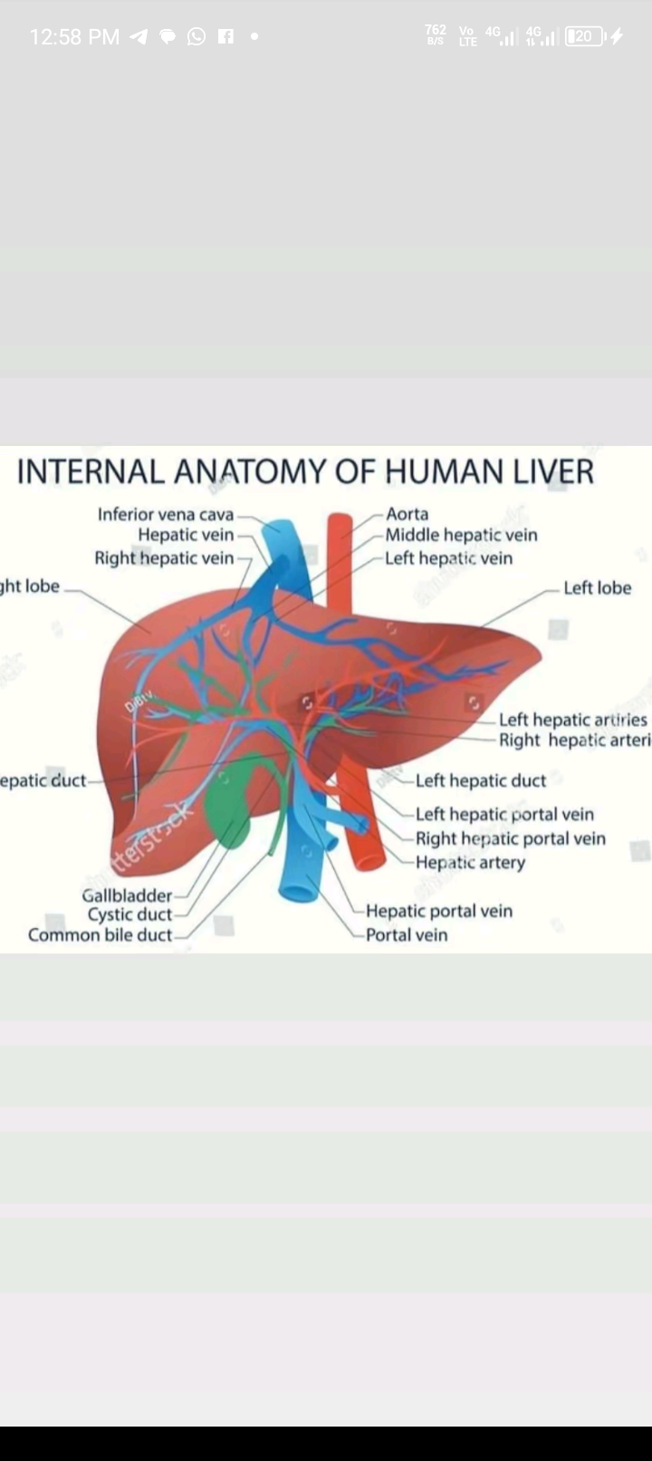
I. Internal Anatomy of the Human Liver
1. Lobes
The liver is divided into anatomical lobes:
Right lobe – the largest; responsible for most metabolic processing.
Left lobe – smaller but essential for digestion and nutrient processing.
Caudate lobe – located on the posterior surface near the inferior vena cava.
Quadrate lobe – situated near the gallbladder.
Clinically, doctors also use functional lobes and segments based on blood supply (Couinaud segments), dividing the liver into eight segments, each with its own vascular inflow and biliary drainage. This segmentation helps surgeons remove diseased portions while preserving healthy tissue.
---
2. Blood Supply
The liver has a unique dual blood supply:
Hepatic artery – delivers oxygen-rich blood from the heart.
Portal vein – brings nutrient-rich blood from the intestines, spleen, and pancreas.
These vessels branch extensively until they reach microscopic structures called sinusoids, where blood is filtered and processed.
---
3. Hepatic Lobules
The liver is composed of millions of hepatic lobules, its functional units. Each lobule:
Is hexagonal in shape.
Contains hepatocytes (liver cells) arranged in plates.
Has a central vein that collects filtered blood.
Includes portal triads at each corner, containing:
a branch of the portal vein
a branch of the hepatic artery
a bile duct
Blood flows inward through sinusoids toward the central vein, while bile travels in the opposite direction, eventually draining into the hepatic ducts and gallbladder.
---
4. Biliary System
The liver produces bile, which helps digest fats. The bile flows through:
Canaliculi → Small ducts → Left and right hepatic ducts
→ Common hepatic duct → Gallbladder or small intestine
Any blockage in these ducts can lead to jaundice or impaired digestion.
---
5. Liver Cells
The liver contains specialized cells, including:
Hepatocytes – perform metabolism, detoxification, protein synthesis.
Kupffer cells – immune cells that remove bacteria and debris.
Stellate cells – store vitamin A; can cause fibrosis if activated by chronic injury.
Endothelial cells – line sinusoids and allow for efficient exchange between blood and hepatocytes.
---
II. How to Protect the Liver
1. Maintain a Balanced Diet
Eat plenty of fruits, vegetables, whole grains, and lean proteins.
Limit added sugars, refined carbs, and trans fats, which contribute to fatty liver disease.
Include liver-friendly foods: leafy greens, nuts, olive oil, berries, and fish rich in omega-3s.
---
2. Limit Alcohol Consumption
Excessive alcohol is a leading cause of liver cirrhosis and inflammation.
General guidance:
Moderate intake: up to 1 drink/day for women and 2 for men (but less is always safer).
People with liver disease should avoid alcohol entirely.
---
3. Avoid Toxins and Unnecessary Supplements
The liver filters chemicals. Overexposure to:
Industrial toxins
Solvents
Certain herbal supplements (kava, comfrey, high-dose green tea extract) can cause injury. Always follow dosage instructions for medications and avoid mixing substances without guidance.
---
4. Maintain a Healthy Weight
Obesity increases the risk of non-alcoholic fatty liver disease (NAFLD).
Regular exercise and weight control significantly reduce fat accumulation in liver cells.
---
5. Practice Safe Medication Use
Never exceed recommended doses of acet